
Overview
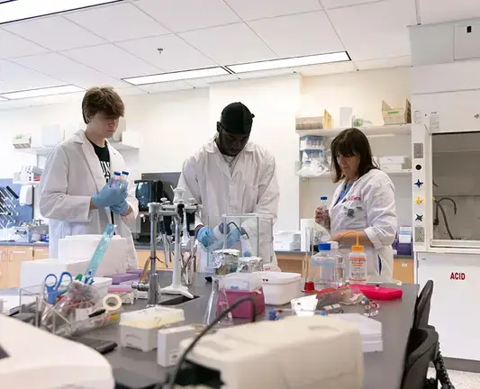
The International Genetically Engineered Machine (iGEM) competition is a multidisciplinary competition in which team members work together to create a solution to an everyday problem. Teams will design, build, test, and measure a system of their own design using interchangeable biological parts and standard molecular biology techniques. They must also consider the human and environmental aspects of their work. The Chemistry and Biochemistry Department oversees the program, but students of any major can become a member of the SUNY Oneonta team.

Grand Jamboree Competition
SUNY Oneonta’s iGEM team competes on an international stage at the iGEM Grand Jamboree, where teams of students from around the world “push the boundaries of synthetic biology by tackling everyday issues facing the world.” Our team has won four consecutive silver medals at the competition and a prestigious gold medal at the 2024 Grand Jamboree in Paris.
Featured News
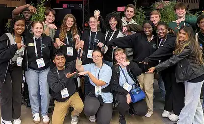
The SUNY Oneonta iGEM (International Genetically Engineered Machine) team returned to Paris in late October to compete in the annual iGEM Grand Jamboree and received a gold medal in its first-ever collaboration with students and faculty from Hartwick College.
Straight From Our Students
I learned the importance of synthetic biology to our world and how one small idea can have a huge impact. I also learned that my love for research is far greater than I could have imagined.
Frequently Asked Questions
The iGEM (International Genetically Engineered Machine) Foundation is a nonprofit organization dedicated to supporting Synthetic Biology. As part of its mission, the iGEM Foundation organizes three initiatives:
- Registry of Standard Biological Parts - An open access repository of standardized DNA parts for use in the creation of devices. These parts are available to academic, nonprofit and community labs.
- iGEM Competition (aka the Giant Jamboree) - An annual synthetic biology event geared toward undergraduate university students, as well as high school and graduate students. Multidisciplinary teams from all over the world work to design, build, and test a biological machine created to address a societal need.
- After iGEM – A mentoring and advocacy program created to support the more than 40,000 iGEM alumni in their future endeavors in synthetic biology and beyond.
For decades now, humans have modified the characteristics of organisms through the use of genetic engineering. Genetic engineers cut and paste together genes from one organism into another to alter the characteristics of a species. Using these recombinant (recombined) DNAs, genetic engineers have created many types of Genetically Modified Organisms (GMOs), from pest resistant crops, bacteria that can clean up oil spills, and yeast that make human insulin.
While effective, traditional genetic engineering is limited in scope, usually involving the transfer of one or a few genes to a new cell. Recombinant DNA technology is also time consuming, expensive, and requires extensive customization to effectively combine genes from one organism with another. Synthetic biology represents a new approach to genetic modification, applying engineering principles to genetic manipulation. In particular, the engineering concepts of standardization, modularity, and abstraction are critical.
Synthetic Biologists view genes, regulatory elements, enzyme target sites, and other DNA features as "parts" and cells as "molecular machines". Parts can be joined to create devices – a collection of parts that creates a function. Many devices can be introduced into a cell (called a chassis) to create a new system, which can allow the cell to complete a new high-level task. This view allows the user to reduce the complexity of building a device and a system, making it easier to understand.
In addition, parts are standardized for assembly into devices by the addition of prefix and suffix sequences. The exact prefix and suffix sequences used form assembly standards, which are used to simplify the process of joining parts. There is even an open source language that has been developed, The Synthetic Biology Open Language (SBOL) that is used to help abstract the design of parts, circuits and systems.
DNA parts in standardized form are most often created in the laboratory synthetically. Information regarding DNA sequences is obtained from online genomic databases such as the National Center for Biotechnology Information (NCBI), maintained by the National Institutes of Health. This information is then used to synthesize DNA parts using artificial gene synthesis technology. Once synthesized, parts can be joined together using common molecular biology techniques such as, cut-and-paste cloning, Gibson assembly, golden gate cloning. Assembled devices are then inserted into a DNA vector, and the assembly is verified. Confirmed device assemblies and then used to transform a target cell, and the function of the device is measured using an appropriate assay. Based on assay results, the constructed device may be subjected to rounds of refinement to improve or modify functions to the required specifications.
To Learn More
Visit SynBioBeta or listen to the SynBioBeta podcast.
Book Review- "Right/Wrong: How Technology Transforms our Ethics" (October 13, 2020)
Synthetic Biology in the News
Food Navigator-usa.com, October 10, 2020: Real Honey, Without the Bees?
Axios, September 30, 2020: The age of engineering life begins
Wired.com: Synthetic Biology
New York Times, May 15, 2019: Scientists Created Bacteria With a Synthetic Genome. Is This Artificial Life?
Students of all majors can participate in iGEM and we are always looking for new members! To learn more about join the team or volunteering to support the team as a mentor, email igem@oneonta.edu.
- Unlike other research projects, students get to investigate and solve a problem of their own choosing.
- The development of practical skills, such as wet and dry lab procedures, designing surveys, web design, and many others.
- Investigating the relationship between science innovation and societal norms and values.
- Gaining experience relevant to courses taken at SUNY Oneonta.
- Experience working as part of a multidisciplinary team to complete tasks.
- Collaboration with peers and experts from all over the world.
- Experience communicating to a diverse audience and receiving feedback from a experts in a variety of fields.
- Participation in job and networking opportunities as part of the project, at the competition, and through the After iGEM alumni program.
Project topics have ranged from "CyanoSpectre" – engineering a cyanophage "toolkit" that other synthetic biologists can use to make it easier to genetically engineer and build beneficial properties into cyanobacteria – to “pHish and CHIPS" – creating a device that would neutralize water automatically after detecting the presence of extreme pH imbalances.
Learn more about previous iGEM teams and their projects on the iGEM Wikimedia pages:
2023 iGEM Team